
Plastic surgeon before and after photos can help explain a proposed change, but they need comparable views, a stated timepoint and an accurate account of the treatment. A striking pair does not establish a complication rate or predict your own result. Use photographs to ask specific questions about what changed and what remains unknown, alongside a clinical consultation. This guide provides an original image-context worksheet, not a provider ranking or an assessment of any individual photograph.
Start by separating three questions: are the images comparable, what care and follow-up do they represent, and how relevant is that example to your situation? Those questions require different information. A pair can be technically consistent but show a different starting problem, while a relevant case can be hard to interpret because the lighting or view changed. Keeping the questions separate makes the consultation more useful.
What does a photograph actually show?
Plastic surgeon before and after photos show the visible appearance captured under particular conditions. They do not directly measure every aspect of function, comfort, sensation, satisfaction or safety. Ask what the image is intended to demonstrate rather than treating a cosmetic impression as evidence about all those outcomes. A photograph can support a discussion of a contour while leaving the patient’s recovery experience unknown.
The 2001 paper Guidelines for Standard Photography in Plastic Surgery discusses the need for consistent, comparable clinical images. It concerns photographic methods of its period, including film equipment, rather than validating every current digital gallery. Its enduring contribution here is the distinction between a permanent visual record and an uncontrolled comparison. This guide does not copy the paper’s figures or claim that its equipment recommendations are a modern universal prescription. 1
Use a precise observation in your own notes. Instead of writing excellent result, identify the visible feature that prompted your question and the view in which it appears. Then ask the clinician what that feature represents and whether another view changes the interpretation. Your note is an observation to discuss, not a diagnosis or a professional assessment of the operation.
Compare the view before interpreting the change
Look for the same region from a similar direction, with similar framing and posture. If one image is closer, rotated or differently cropped, ask for a comparable pair. Avoid attempting to correct a clinical image yourself and calling the result an objective comparison. Resizing or rotating a screenshot can introduce its own uncertainty, particularly when the original capture information is missing.
For plastic surgeon before and after photos, write down what you can see and what you cannot establish. A front-facing pair may help with one visible feature while leaving side contour or scar location outside the frame. A close-up may omit surrounding context. Do not infer a complete result from the one view selected for a promotional post.
Ask whether a fuller set of standard views is available for discussion within the practice’s consent and privacy arrangements. The purpose is to understand the example, not to demand access to another patient’s complete medical record. If the relevant view is unavailable, mark that limitation rather than filling the gap with an imagined outcome.
A measured example of photographic inconsistency
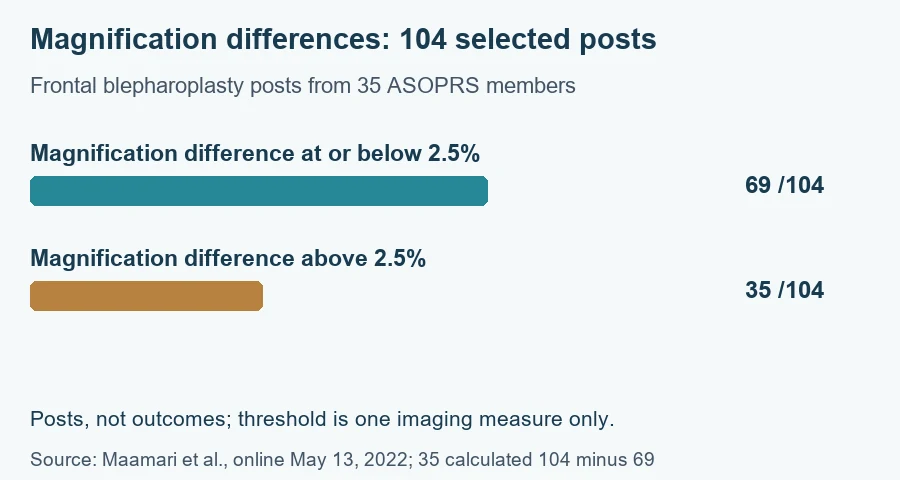
A 2022 original study analyzed 104 frontal blepharoplasty posts from 35 ASOPRS members on Instagram over a four-month collection period. Its abstract reports that 69 of the 104 photosets had a magnification difference of 2.5% or less. Subtracting 69 from 104 gives 35 above that threshold in this selected sample. The threshold concerns one imaging measure; it is not a success rate or a universal standard for judging a surgeon. 2
The original chart accompanying this guide plots those two magnification groups with the same denominator. It does not combine the study’s other imaging metrics into a quality score. Posts are the unit, not unique surgical patients or procedures with a measured outcome. The collection was a particular specialty, platform and sample, so these counts cannot establish how often all plastic-surgery photographs are misleading.
The useful lesson for plastic surgeon before and after photos is to ask about comparability before attributing every visible difference to treatment. The study identifies imaging variation, not the intent behind a particular post. A mismatch can be a reason for clarification without establishing deception, malpractice or a poor clinical result. Keep that distinction in your notes and in any conversation about the images.
| Original chart category | Posts in the selected sample | Interpretation boundary |
|---|---|---|
| Magnification difference at or below 2.5% | 69 of 104, reported | One imaging metric, not proof that every other condition matched |
| Magnification difference above 2.5% | 35 of 104, calculated as 104 minus 69 | Not a count of failed operations or dishonest providers |
| Study scope | Frontal blepharoplasty posts from 35 ASOPRS members | Selected social-media sample, not national prevalence |
Light, color and image processing
Lighting can alter what a surface or contour appears to show. Ask whether the capture conditions were consistent rather than interpreting a brighter image as better skin or a deeper shadow as a larger anatomical change. The 2022 study assessed luminosity, color and edge-related measures as well as magnification. This guide uses only the stated magnification groups in its chart; it does not infer all-image quality from them. 2
For plastic surgeon before and after photos, ask whether any processing changed the appearance of the treated area. Cropping for display and altering a visible treatment feature are different operations, but a viewer may not know what occurred from the final image alone. Request an explanation rather than claiming that a filter can be diagnosed reliably by eye.
Be cautious about comparing a screenshot from one platform with an original image shown elsewhere. Compression, display settings and reproduction can add differences unrelated to the treatment. A practice’s explanation of the original record is more useful than a confident conclusion based on two files collected under different conditions. The worksheet should identify the source you actually viewed.
Timepoint belongs in the comparison
An after image needs a stated interval from treatment. A photograph shortly after a procedure and a photograph months later answer different questions about the visible course. Ask the clinician what is expected to change over time in the relevant procedure and what the displayed timepoint can reasonably illustrate. This guide does not assign a universal final-result date across operations.
Plastic surgeon before and after photos should be interpreted with the actual follow-up context, not an assumed timeline. If a caption says after without a date or interval, write timepoint unknown. Do not assume the most favorable image is the final one or that a later photograph establishes permanent stability. Ask what later information is available and what remains uncertain.
If different examples use different intervals, avoid arranging them as if they were directly comparable endpoints. You can still discuss each case, but the timepoint difference belongs in the record. An original gallery worksheet should have a separate field for treatment date or interval rather than hiding it in a general notes column. For checking the actual licensing-board record, see plastic surgeon disciplinary history.
Identify the treatment represented
For plastic surgeon before and after photos, ask which procedure or combination of treatments produced the displayed example. A change after multiple interventions does not isolate the effect of one. A caption with a broad procedure name may omit details important to the comparison. The clinician can explain the actual scope without promising that the same combination is appropriate for you.
Keep surgical treatment, nonsurgical treatment and other changes distinct when the information is available. Do not infer a particular technique from an image or assume that every difference came from the named operation. Weight change, additional care and timing may be relevant questions for the case discussion, but they should not be invented as facts about the photographed patient.
The ASPS patient consultation questionnaire includes asking to see before-and-after photographs for the procedure and asking what results are reasonable for the individual. Those are paired questions: the example starts the conversation, and the clinician relates it to your actual assessment. The questionnaire is an educational checklist, not an endorsement of any displayed case or an outcome guarantee. 4
Match the example to the question you are asking
Similar age or appearance does not establish identical anatomy, treatment needs or response. For plastic surgeon before and after photos, ask what part of an example is relevant to your concern and what part is not. The useful comparison may concern a specific tradeoff, such as visible contour versus scar placement, rather than an overall resemblance to the photographed person.
Make a short list of features you want to discuss without demanding an exact replica of someone else’s result. A clinician can explain what the proposed plan aims to change, what it cannot establish and which alternatives merit discussion. Keep the photograph as a communication aid rather than a specification that overrides your own clinical assessment.
If you like one feature but dislike another in a displayed case, record both. Ask how the relevant tradeoff is considered in planning. A gallery can be useful precisely because it raises those questions, not because every visible feature looks ideal. This is a more informative conversation than assigning the entire case a star rating.
| Context field | Question to ask | If unavailable |
|---|---|---|
| View and framing | Are the same region and direction shown? | Record the comparison limit |
| Treatment scope | Which actual procedures are represented? | Do not attribute the change to one unconfirmed technique |
| Timepoint | How long after treatment was the image taken? | Mark interval unknown |
| Personal relevance | What is and is not comparable to my assessment? | Ask during the clinical consultation |
A gallery does not supply an outcome denominator
Plastic surgeon before and after photos usually present selected examples. The number shown does not tell you how many total patients were treated, how cases were selected or how many experienced a complication. Do not calculate a success rate from the proportion of attractive photographs in a gallery. Ask for the appropriate evidence and explanation when your question concerns risk or expected outcomes.
Similarly, a large gallery does not verify credentials, facility arrangements or the management of complications. Those are separate questions with separate evidence. A small gallery may reflect consent or display decisions rather than a simple measure of experience. Avoid treating either size as a shortcut to a provider ranking. For checking the exact specialty-board credential, see plastic surgeon board certification.
Use a visible example to ask about limitations as well as benefits. Ask what a photograph cannot show about the course of care and how the team discusses dissatisfaction or further treatment. This guide does not provide a practice’s revision policy; it helps identify which missing information should be obtained from that practice’s actual process.
Photography consent and comfort
A 2024 original survey recruited 411 respondents through a crowdsourcing platform to examine comfort with clinical photography scenarios. The sample was predominantly White and many respondents had no prior plastic-surgery experience. Its findings concern reported comfort under the described scenarios, not a nationwide legal-consent rule or evidence that one device guarantees confidentiality. The actual publication was online March 14, 2024, with an August 2024 issue. 3 For assessing the limits of online-review evidence, see plastic surgery online reviews.
When your own plastic surgeon before and after photos are discussed, ask about the purpose of capture, storage, access and any proposed use outside your care. A clinical record and public promotional display are different contexts to understand. Read the actual consent materials and ask about unclear terms rather than assuming that agreement to care answers every image-use question.
You can also ask who will take the images and how privacy and comfort are handled during the session. This guide does not determine an individual consent dispute or promise a specific device policy. It encourages a factual conversation about the process before sensitive photographs are taken or shared.
An original photo-to-question worksheet
For each set of plastic surgeon before and after photos you discuss, record the source, visible feature, known view, treatment scope, timepoint and unanswered question. Use the practice’s own case identifier if one is available, without copying identifying patient information into an unnecessary personal file. A short description can often help you locate the example during the consultation.
Keep observation and interpretation in different columns. Observation might say side view or interval not stated. Interpretation might be a question about what the change represents. Do not enter the interpretation as a verified clinical fact before the clinician explains it. That distinction makes the worksheet useful even when the example is incomplete.
| Worksheet entry | Write what is actually known | Next question |
|---|---|---|
| Example source | Gallery or case identifier and date viewed | Can we discuss the actual record? |
| Visible feature | Specific region and view | What does this feature demonstrate? |
| Context gap | Missing scope, interval or comparability fact | What information would clarify it? |
| Clinical discussion | Actual explanation and remaining limits | How does this relate to my assessment? |
Track explanations when several images are shown
A consultation may move quickly through several plastic surgeon before and after photos. Give each example a short, nonidentifying description so an explanation about one case is not accidentally applied to another. If the clinician explains that one photograph shows an early timepoint and another shows later follow-up, preserve that difference rather than summarizing both as final results.
When you return to the gallery later, check that you are looking at the same example and view. A cropped social-media post and a fuller consultation set can be different displays of a case. Ask about the actual context if they seem inconsistent. Do not infer that a missing image proves the outcome changed or that a new caption supplies all the facts absent from the earlier one.
The worksheet can finish with two separate entries: the feature the example helped explain and the question it could not answer. That division keeps a useful visual conversation from expanding into an unsupported conclusion about safety, durability or your own likely response.
Use images alongside the full consultation
Plastic surgeon before and after photos are most useful when they make a question more specific. Bring the worksheet to the clinical discussion and ask for an explanation of the relevant example. Keep the resulting answer within its stated scope. A discussion of one visible feature does not establish your candidacy, a precise outcome or freedom from complications.
The original chart and worksheet in this guide contain no patient photographs and no simulated surgical results. They explain a reported imaging measure and a way to organize questions. A conceptual illustration, a computer preview and a real patient outcome image have different roles; ask which role applies whenever a visual is shown to you.
Your final notes should identify what the example helped explain and what still needs other evidence. That is a useful outcome of reviewing photographs. It supports a more informed consultation without converting selected visual examples into a promise about the operation you may be considering.
Sources and their limits
Original author abstracts and the ASPS questionnaire were inspected October 12, 2026. The photography-method paper is a historical 2001 source; the quantitative analysis is a selected 2022 social-media sample; the 2024 survey concerns reported comfort. No named provider or clinical outcome photograph is republished here. The graphic is newly drawn from the stated sample counts, not copied from a journal.
| Source | What it supports | Important boundary |
|---|---|---|
| R01: Standard photography guidance, March 2001 | Consistent and comparable clinical photography | Historical equipment context, not current universal specifications |
| R02: Oculoplastic social-media photography, online May 13, 2022 | Actual 104-post magnification sample | Not surgical success, deception prevalence or national risk |
| R03: Photography comfort survey, online March 14, 2024 | Reported comfort in surveyed scenarios | Crowdsourced respondents, not a universal consent rule |
| R04: ASPS consultation question checklist | Examples and individual expectations discussed together | Undated questionnaire, not provider endorsement |
Sources and review scope
Plastic Surgery Editorial Team. Source records checked through October 12, 2026. Review scope: source and editorial checks for general education. No individual clinical assessment is represented.
- Guidelines for standard photography in plastic surgery. pubmed.ncbi.nlm.nih.gov. Source date: 2001-03. Checked 2026-10-12.
- Quantitative analysis of oculoplastic social-media photographs. pubmed.ncbi.nlm.nih.gov. Source date: 2022-05-13. Checked 2026-10-12.
- Patient comfort with before and after photography. pubmed.ncbi.nlm.nih.gov. Source date: 2024-03-14. Checked 2026-10-12.
- ASPS consultation question checklist. plasticsurgery.org. Publication/update date not stated. Checked 2026-10-12.