
Split earlobe repair addresses an acquired cleft or elongated piercing defect in the soft earlobe. A useful plan separates three goals: closing the defect, restoring the lower contour and deciding whether a piercing channel should remain. These goals can lead to different proposals. An enlarged hole with tissue still beneath it is not the same starting problem as a complete split, tissue loss or a previously repaired cleft.
Published techniques offer several ways to approach those problems, but a favorable small series does not guarantee an invisible scar, permanent strength or immediate return to earrings. This guide explains the decisions and reads original reports with their actual participants, repaired sites and follow-up. It does not provide a repair technique, an individual diagnosis or instructions for treating a fresh injury.
First distinguish a settled defect from a current injury
An old split being considered for elective repair is a different situation from recent trauma, bleeding or suspected infection. Do not postpone assessment of a new injury while choosing a cosmetic technique online. If you suspect an infected piercing, obtain prompt medical help. NHS guidance identifies features such as painful hot swelling, pus or feeling unwell as reasons to seek assessment; it is general piercing guidance rather than a diagnosis of any particular earlobe. R01
Describe the history before focusing on appearance. Was there an abrupt snag, a slowly lengthening hole, an intentionally stretched piercing or an earlier repair? Is your concern the opening itself, the lower edge, the overall lobe size or something else? Give the clinician the history of prior treatment and any current symptoms. These details help define the problem being evaluated; they are not a self-assessment score for candidacy.
A contour concern involving the wider ear also deserves its own description. Closing a lobe defect should not be assumed to change how far the upper ear projects from the head. Likewise, reducing an elongated lobe is a different goal from closing a split. Keep each requested change visible so that an attractive procedure label does not conceal a mismatch between the treatment and your concern.
Document the remaining tissue and the lower edge
Original acquired-cleft research distinguishes incomplete from complete splits and considers the lower border, tissue loss and previous recurrence. Those features matter because a closure must be planned around the actual tissue that remains. The 2020 composite-technique report explicitly organized its assessment around these differences. Its classification is an author’s planning framework, not a home examination or a universally mandatory grading system. R02
Ask the clinician to describe your starting defect in plain language. If a bridge of tissue remains below the opening, ask how that feature affects the proposed repair. If the lower edge is already uneven or tissue is missing, ask what can be restored and what limitations remain. If there was previous surgery, ask how the existing scar changes the plan.
Document each ear independently. A complete split on one side and a stretched hole on the other may call for different treatment, even if the desired appearance is similar. Identical procedure names do not establish identical tissue problems. The estimate and consent should identify which side is being treated and what each treatment is intended to accomplish.
| Starting concern | Feature to clarify in examination | Decision that remains separate |
|---|---|---|
| Elongated opening with tissue below | Opening and remaining lower tissue | Whether a channel should remain after repair |
| Complete split | Separated edges and available tissue | Which lower contour can reasonably be restored |
| Tissue loss or a retracted edge | Amount and condition of remaining tissue | Whether a more involved reconstruction is proposed |
| Previous repair that split again | Prior technique, scar and new injury history | Why a revised plan would differ |
| Different defects on the two sides | Each side’s description and intended change | Whether both need treatment at the same visit |
Do not let a photograph supply missing information
A photograph may help describe a visible concern, but it cannot establish tissue quality, sensation or the complete history of a repair. If you compare examples, ask whether the example began with a similar defect and whether the treatment included additional contour work. A healed image is evidence of the appearance pictured, not proof that the person had no complications or could safely wear a particular earring.
Keep a record of the feature you want changed and the feature you want retained. If you like the lobe’s overall size but dislike the split, say so. If contour is more important to you than keeping the original piercing, make that priority explicit. This allows a proposal to be assessed against your preference rather than against someone else’s photograph.
Decide what should happen to the piercing channel
Some published repairs aim to retain or recreate a channel, while others close the opening and leave later piercing as a separate decision. The 2024 original comparative report studied two channel-preserving approaches. Its existence shows that channel management is part of the treatment question; it does not establish that retaining a channel is appropriate for every defect. R03
Ask whether the proposed result is a closed lobe, a retained opening or a newly formed opening. Then ask where that opening would be relative to the lower edge and existing scar. “You can wear earrings afterward” leaves too much unexplained if wearing earrings is a principal reason for seeking repair.
Keeping an opening is not permission to load it immediately. A repair plan, a healing assessment and a return-to-jewelry decision answer different questions. Obtain the team’s instructions for when and how piercing or earring use should be considered. Do not select a timetable from another technique’s published postoperative routine.
Treat later piercing as a planned decision
If the opening will be closed, ask who will assess readiness for another piercing, how its location will be chosen, and whether that service is included in the quote. If an opening will remain, ask what follow-up is needed before using it. In either case, document the advice rather than assuming that a visible channel means the repair is ready for ordinary jewelry.
Your future preferences also matter. If you no longer want a piercing, channel preservation may not be a goal worth pursuing. If you want earrings only for particular occasions, describe that pattern without treating it as proof of safe loading. The clinician can address the intended use within a plan matched to the repaired tissue.
Read the 2020 series using people and lobes separately
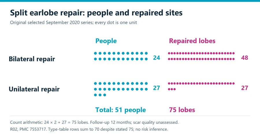
The original September 13, 2020 report describes 75 repaired lobes assessed at 12 months. It states that 24 people had bilateral repairs and 27 had unilateral repairs, all women. Those descriptions correspond to 51 people and 75 lobes: 24 times two plus 27. The person count is a transparent calculation from the reported treatment groups, not a separately quoted enrollment number. R02
A repaired lobe is not an independent person. Site-specific contour observations may appropriately be counted by lobe, while a person’s overall experience is a different unit. Two repaired sides can share the same person’s healing circumstances and preferences. Do not turn a site count into a count of unrelated participants when interpreting a report.
The paper also contains a denominator caveat. Its displayed type rows contain seven, 30 and 33 cases, which add to 70, although the table states a total of 75. The header notes an excluded subtype, but the displayed material does not reconcile the difference into a clearly counted additional group. This guide does not invent five missing cases or recalculate a technique-specific complication rate from that table.
The supporting chart shows the reported unilateral and bilateral groups as people and their corresponding repaired lobes. It makes the arithmetic visible without implying an outcome probability. The study assessed selected contour findings and recurrence, but explicitly did not assess scar quality. A reader whose main concern is a discreet scar therefore needs a separate discussion; the presence of a 12-month follow-up does not mean every desired endpoint was measured.
Ask what a smaller comparative study establishes
The original comparative article published online June 8, 2024 and assigned to the April 2025 issue included 12 people. It compared composite closure and double-flap treatment and assessed participants on postoperative days 14 and 90. The accessible publisher abstract reported favorable results with both approaches. This is a small, short-term comparison, not proof of multiyear durability or superiority for every cleft type. R03
A technique may have an appealing name or a sensible rationale without having strong comparative evidence for your particular endpoint. Ask what comparable cases inform the recommendation and how the desired feature was assessed. If the available evidence is a technical description or small case series, that limitation should be visible rather than replaced by a claim of universal success.
| Evidence question | Detail to look for | Avoid this shortcut |
|---|---|---|
| How many were treated? | People, repaired lobes and any bilateral treatment | Calling every treated side an unrelated participant |
| What defect was studied? | Complete, incomplete, recurrent or tissue-loss cases | Applying a favorable report to every starting problem |
| What was measured? | Lower contour, recurrence, scar quality or another defined endpoint | Treating an unmeasured outcome as normal |
| When was it assessed? | Actual follow-up point or range | Making a short-term observation a permanent guarantee |
| Was there a comparison? | Defined comparator and actual group allocation | Declaring superiority from an uncontrolled series |
The appropriate use of this research is to improve the questions you ask. It does not allow a reader to choose an incision pattern, a suture material or a reconstructive flap. Those are operative decisions requiring examination and qualified treatment.
Make scars and contour explicit goals
Closing a defect and restoring the lower outline are related but distinct endpoints. A repair can close while a contour concern remains. Similarly, a photograph can look satisfactory while the person has a concern about the scar’s feel or visibility. Define which outcome matters most and how it will be assessed.
An ASPS-hosted clinician explanation describes earlobe repair as requiring evaluation of the opening and surrounding anatomy and notes that a scar remains after healing. It represents that author’s clinical explanation, not a national outcome study or a guarantee that every scar will be fine or invisible. R04
Ask the treating team to explain how your scar history and existing tissue affect the discussion. Bring information about earlier healing problems or repairs if applicable. Do not assume that an ordinary-looking scar in a promotional photograph predicts your own result. A useful explanation should acknowledge uncertainty while showing how the proposed assessment and follow-up address your concern.
If you are judging photographs, keep framing and presentation consistent. Earrings can conceal part of the lobe, so an image with jewelry may not answer a question about the lower edge or repair line. Ask for an explanation of what the image shows and what it does not. A visual comparison is most useful when it is linked to a defined feature rather than an overall impression of perfection.
Organize follow-up around the repaired ear
Before treatment, obtain the instructions for wound care, dressings, medication if prescribed, follow-up and the changes that require advice. Ask how the plan accounts for everyday items that contact or pull on the ear, including clothing, headphones or your usual accessories. These are practical questions for the team, not permission from this guide to use a particular item at a particular time.
If you work in a role involving ear-mounted equipment or a risk of snagging, describe the actual tasks. Returning to work is a different question from readiness for ordinary jewelry, another piercing or a final appearance assessment. A broad claim of next-day recovery may not answer the particular activity you need to perform.
Keep the contact instructions accessible and ask what follow-up is arranged if you live far away. A brief procedure still needs an assessment plan. If a new symptom worries you, contact the treating team or obtain urgent care according to its seriousness; do not use a general article to decide that a change is harmless.
The recovery planning guide helps organize practical support and follow-up questions. The consultation question guide can help document the proposed setting and qualifications. Neither replaces instructions for your repaired ear.
Prepare a separate record for a recurrent split
If an earlier repair has opened again, do not assume that the original method was unsuitable or that an allergy caused the recurrence. Start with the known sequence: when treatment occurred, whether the opening healed, when jewelry or other contact resumed, and whether there was a definite new injury. Mark uncertain details as uncertain. A remembered event is useful history, but it is not a demonstrated cause.
Ask for available records of the previous procedure and subsequent assessments. Photographs from before and after the earlier repair may help describe the changes, provided their dates and context are known. They cannot substitute for examination of the current defect. A clinician reviewing recurrence needs a record of what happened, rather than a reconstruction based solely on how the scar looks now.
The new proposal should explain what would be different and why. That may concern the present tissue problem, the intended channel or the lower contour; the answer needs to be specific to the examination. Ask which limitations from the previous repair remain and which outcome the new plan seeks to improve. Do not interpret the existence of a more elaborate technique as proof of greater durability.
Finally, agree on how the result will be reviewed. If your main concern is another split during ordinary use, an early closed wound alone does not answer that question. Keep the use instructions and longer follow-up discussion connected to the goal you brought.
Compare scope before comparing a price
A quote should identify one or both ears, the defect being treated, any contour reconstruction, channel management, the treatment setting and included follow-up. If later piercing is a separate service, record that separately. An estimate for closing one uncomplicated defect cannot be treated as the same service as a bilateral or recurrent reconstruction.
This guide does not adopt an old blog range as a current national price. Ask for a written estimate and confirm which professional, facility, medication or additional-care expenses apply. If insurance might be involved, establish the documented reason and coverage with the relevant parties rather than assuming that the word repair guarantees payment.
| Quote item | What to record | Why it helps comparison |
|---|---|---|
| Side and starting defect | Each ear and planned component | Prevents unlike scopes from appearing equivalent |
| Channel management | Closed, retained or recreated opening | Makes later piercing arrangements visible |
| Follow-up | Included assessments and separate charges | Distinguishes procedure fee from care scope |
| Additional treatment | Assessment and fee policies | Does not confuse a financial policy with clinical suitability |
| Financing or insurance | Total terms or confirmed coverage | Avoids treating a monthly payment as the total expense |
Use the cost planning guide for organizing a complete estimate. Keep financial and clinical questions separate: a low price does not define the best reconstruction, and a free revision clause does not establish that another operation would be appropriate.
Leave with a plan that answers your actual goal
The decision record should describe the starting defect, the desired lower contour, the intended piercing arrangement, the evidence’s relevant population and the care plan. It should also identify what remains uncertain, especially if your principal concern was not measured in the studies being cited.
If the proposed operation solves a different problem from the one that matters to you, seek clarification before proceeding. You can decide to close a split without retaining a piercing, prioritize contour over the original hole location, or wait while considering the tradeoffs. Repair is most useful to understand as a set of tissue and use decisions, supported by examination, rather than a promise that an ear will become indistinguishable from one that was never injured.
Sources and review scope
Plastic Surgery Editorial Team. Source records checked through October 12, 2026. Review scope: source and editorial checks for general education. No individual clinical assessment is represented.
- NHS infected piercing guidance. nhs.uk. Source date: 2023-04-18. Checked 2026-10-12.
- Original acquired split-earlobe classification and composite-repair series. pmc.ncbi.nlm.nih.gov. Source date: 2020-09-13. Checked 2026-10-12.
- Original comparison of composite and double-flap repair of traumatic split earlobes. link.springer.com. Source date: 2024-06-08. Checked 2026-10-12.
- ASPS-hosted clinician explanation of earlobe repair. plasticsurgery.org. Source date: 2021-08-11. Checked 2026-10-12.