
Ptosis surgery raises a drooping upper eyelid by addressing its lifting mechanism. It is different from removing excess eyelid skin, although both concerns can exist together. The useful decision begins with an eye assessment: why the lid is low, how it affects vision, how its lifting muscle works, and whether the eye can be protected after correction. A higher lid is not the only goal; eyelid closure, comfort and the relationship between the two sides also matter.
A suddenly drooping eyelid needs urgent medical assessment rather than an elective surgery consultation. NHS guidance identifies sudden droop and changes in eyesight among reasons to seek urgent care. Sudden inability to see or sudden severe eye pain requires emergency assessment. Use the appropriate urgent or emergency service where you live rather than waiting for a cosmetic appointment. NHS eyelid symptom guidance NHS vision-loss guidance
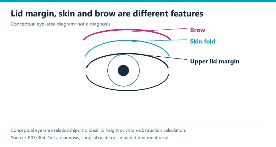
Identify which part of the upper eye area is changing
Ptosis refers to a low upper eyelid margin, the edge nearest the eye. Excess skin can hang over the eyelid without the same problem in the lifting mechanism. Brow position is another feature. Describing everything as hooded eyes can conceal these distinctions and make it difficult to understand what a proposed operation is intended to change. Ask the examiner to show you which feature or combination of features is relevant.
Cambridge University Hospitals explains that ptosis can obstruct the upper field of vision when the lid margin covers part of the pupil. Its assessment includes the amount of droop, eyelid muscle function and examination of the eyes, with visual-field testing where appropriate. This is more than choosing the desired appearance from a photograph. The findings help determine whether surgery is appropriate and what form of correction might be considered. CUH adult ptosis assessment
NHS guidance describes blepharoplasty as removing excess skin or fat from the eyelids. That distinction matters if you are comparing proposals: a skin-removal procedure is not automatically a repair of a low lid margin. Conversely, raising the margin may leave a skin concern to discuss separately. Confirm the actual scope of each proposed intervention rather than assuming that an eyelid operation includes every feature around the eye. NHS blepharoplasty definition
| Feature | What the assessment distinguishes | Planning question |
|---|---|---|
| Upper eyelid margin | Position of the lid edge and lifting function | Is this the feature the proposed repair changes? |
| Excess eyelid skin | Skin overhang separate from the margin | Is skin removal included or a separate decision? |
| Brow position | Brow height and its relationship to the eyelid | Would the proposed eyelid repair leave this concern unchanged? |
| Eye surface and closure | Ability to protect the eye | How will closure and comfort influence the target position? |
| Difference between sides | Margin, contour and baseline asymmetry | How will both sides be assessed even if only one is treated? |
The table is an organizer for a discussion, not a way to diagnose yourself. Do not measure a photograph and assign a surgical target from it. Expression, gaze and the way the image was taken can alter the visible relationship between features. A clinician needs to examine the relevant structures and function. Ask for an explanation in plain language that connects those findings to the intervention being proposed.
Bring a useful history to the eye assessment
Record when the droop began, whether it involves one or both eyes, and whether it varies. Identify previous eye operations, injuries, contact-lens use and relevant medical conditions. Explain any change in vision or other symptoms. If an old photograph helps establish that the position has changed, ask whether to bring it. A dated history is more informative than an assumption that all adult droop is simply aging.
Guy’s and St Thomas’ describes several possible acquired causes, including changes involving the eyelid tendon after age-related change, contact-lens use, trauma or eye surgery. It also identifies less common muscle and nerve conditions. Those possibilities explain why the cause needs assessment before choosing an elective repair; they do not mean that a reader can infer a diagnosis from the pattern of droop. Guy’s and St Thomas’ ptosis education
Explain the situations in which the concern affects you. Do you notice an upper-field limitation, use your forehead to raise the area, or mainly dislike the resting appearance? These are questions to discuss, not proof of a particular cause or entitlement to treatment. Separating functional and appearance priorities helps the examiner explain which outcomes are realistic and which findings need further investigation.
This guide concerns adult planning. A child’s drooping eyelid needs an appropriate pediatric eye assessment rather than an adult cosmetic plan. Alder Hey explains that congenital ptosis can affect visual development, depending on its extent. Adult study findings and adult recovery arrangements should not be transferred to a child simply because the condition has the same name. Alder Hey pediatric scope
Understand what the measurements are for
Ask the examiner to explain the purpose of the measurements being taken. Lid position and lifting function describe different aspects of the problem. A visual-field test addresses what you can see under the test conditions. The examination also considers the eye itself. These pieces of information support a treatment decision together; one number should not silently become a complete readiness assessment.
If a test is performed with the eyelid in different positions, ask what the comparison is intended to demonstrate and how it will be documented. Do not tape your eyelids at home to reproduce a clinic test or decide that you qualify for an operation. The test method, examination and interpretation matter. Keep a copy of the relevant report if it is being used for a treatment or payment decision.
Questions about treatment need to remain distinct from questions about coverage. An insurer or public service may use specific documentation requirements, while the clinical plan still depends on your individual assessment. A test result that meets a funding criterion does not guarantee a preferred cosmetic result, and a concern that matters to you does not automatically meet every payer’s rules. Ask which decision the test is supporting.
A testing study is not a surgical success rate
A 2023 original pilot study compared virtual-reality superior visual-field testing with standard automated perimetry in 78 eyes from 41 people undergoing functional upper-eyelid assessment. Participants included people with ptosis, brow ptosis and excess skin. Its endpoint was how the tests identified fulfillment of a specified insurance-coverage criterion, not how well an operation corrected the eyelid afterward. Original visual-field testing pilot
Eyes and people are different research units
The study’s 78 eyes are not 78 independent people, and its mixed assessment population is not a pure postoperative ptosis cohort. Reported sensitivity and specificity describe the performance of one test compared with another under that study’s definition. They should not be displayed as the chance of successful ptosis surgery, improved appearance or permanent symptom relief. The endpoint determines what a percentage means.
The paper called for further work on reliability, waiting and testing time, and satisfaction. That is a useful reminder that a promising assessment method does not answer every practical question. When a newer device is proposed, ask how it fits into your eye examination and what the result will be used for. A headset or an automated report should not replace an explanation of the findings and their clinical relevance.
| Evidence claim | Correct question | Meaning to avoid |
|---|---|---|
| A visual-field test identifies a funding criterion | How did it compare with the specified reference test? | A surgical success rate |
| A lid-position measurement changes | What measurement, conditions and timepoint were used? | Automatic proof of comfortable closure or satisfaction |
| An appearance photograph looks improved | Which feature changed and when was the image taken? | A complete functional assessment |
| A patient is satisfied | What question or measure was used? | Proof that every clinical endpoint is normal |
| A clinic quotes a favorable outcome percentage | What population, definition and follow-up produced it? | A personal guarantee for your proposed repair |
If a percentage is used in the consultation, request its definition and observation period. A good position at an early visit is different from longer-term persistence, and a population average cannot settle your preferred balance between appearance, field of view and comfort. Evidence is most helpful when it supports the actual decision rather than when its number is detached from the endpoint that produced it.
For example, a report might describe a satisfactory lid position without explaining whether participants still needed additional treatment for dryness. That would leave the comfort question unanswered even if its positional endpoint were clear. Another report might record improvement on a visual-field test without asking whether the patient was pleased with the contour. Neither result is automatically wrong; each needs to be read as the specific outcome it measured. Use that distinction when deciding what information is still missing from a proposed plan.
The same principle applies to photographs. A close view may show the upper eyelid margin clearly but omit the brow; a wider view may show symmetry without enough detail to assess the margin. Ask which photographs and examination findings will serve as the baseline for each goal. If the result is later discussed using a different view or expression, identify that difference before interpreting it as a clinical change. Image consistency improves communication, but photographs still do not replace the eye examination or functional testing.
A practical outcome record can therefore retain several separate observations: the examined lid position, the visual-field finding if tested, the clinician’s assessment of closure and your report of comfort or appearance. Keeping the observations separate prevents one favorable feature from being used to imply that every other feature has been assessed and resolved.
Balance lid height with eye protection
CUH explains that tightening or shortening the lifting mechanism can make the eye harder to close and expose it to dryness. The desired height must therefore be considered alongside eye protection. Its guidance also discusses overcorrection, undercorrection, contour differences and recurrence. A proposal to raise the lid as far as possible is not a sufficient explanation of the intended result.
Ask how existing dry-eye symptoms or difficulty closing the eye will be assessed and how they affect the plan. Describe the products and treatments you already use for your eyes, including contact lenses, and bring an accurate medication list. This guide does not recommend changing medication, beginning drops or adjusting a prescribed regimen. The treating team should provide individual instructions and coordinate any needed changes with the relevant prescriber.
The two sides also need to be considered together. CUH notes that correction on one side can reveal a tendency for the opposite side to droop. Ask how the examiner has assessed both eyes and how a later change on the other side would be managed. Do not interpret every postoperative difference as an automatic need for another operation; it should be assessed in context with the agreed goals and eye health.
Ask why a particular repair is proposed
The operation should be explained in relation to the assessed cause and lifting function. Ask whether the approach is from the front of the eyelid or its inner surface, what structure is being adjusted, and whether skin removal is a separate component. You do not need technical steps for performing the repair. You need enough understanding to connect the proposed procedure with your own findings and consent.
Clarify the planned setting and anesthesia, including whether participation during adjustment is anticipated. NHS adult information describes local anesthesia as common, but the actual arrangement must be confirmed with your team. Discuss any concern about remaining awake before the day of surgery. A booking described as an eyelid repair should specify the operation, the side or sides and the planned care rather than rely on a generic procedure label.
If more than one procedure is proposed, ask for a separate explanation of each goal. Raising the lid margin and removing skin may be combined in a plan, but they remain different interventions. Identify which feature will be evaluated for each component at follow-up. That makes it possible to discuss an unresolved concern later without confusing it with another part of the operation.
Make postoperative eye care practical
Ask for written instructions about the wound, prescribed eye products, follow-up and activities. The NHS sources describe individualized arrangements for ointment or drops, dressings and review. Those arrangements differ between services, so this guide does not reproduce a universal driving date, contact-lens interval or exercise schedule. Follow the instructions for your actual procedure and clarify anything that conflicts with your ordinary eye care.
Think through tasks that depend on clear vision. Will someone take you home, help with reading instructions or handle transport to a review? Can you manage the prescribed products correctly, or do you need assistance? Ask the team to explain the order and use of medicines in a format you can read. These practical details matter even when an operation is described as a day-case procedure.
The Guy’s and St Thomas’ leaflet advises immediate assessment after surgery if pain increases, vision worsens, redness increases or excess discharge develops. Vision loss should never be left for a routine appointment. Confirm the treating team’s urgent contact route and the emergency service available where you will recover. A follow-up booking is not a substitute for access when an unexpected concern occurs.
Ask how the team distinguishes early swelling from a later position or contour assessment. Keep the review dates and the agreed outcome questions together. If additional treatment is being considered, request the reason, the remaining uncertainty and the intended endpoint. A decision about revision should arise from assessment rather than a casual comparison with an immediately postoperative photograph.
Clarify the financial scope and coverage
The checked sources do not establish a contemporary US national total price for an individualized ptosis repair. UK public-service descriptions also do not determine a US insurer’s decision. Obtain a written estimate for the actual operation and ask the payer about its current documentation requirements. A cosmetic blepharoplasty price should not be relabeled as a ptosis price without checking which services are included.
| Financial or consent item | Detail to request | Why it matters |
|---|---|---|
| Operation and sides | Named repair on one or both eyelids | Prevents a generic eyelid fee from concealing scope |
| Additional skin procedure | Separate clinical goal and billing status | Distinguishes margin correction from skin removal |
| Examination and testing | Included services and payer requirements | Keeps funding evidence separate from an outcome promise |
| Facility and anesthesia | Responsible services and separate charges | Defines the full planned cost |
| Later review or revision | Included care and written financial policy | Makes the possible commitment beyond initial surgery visible |
Compare written scopes rather than only the headline quote. Include the help and transport you may need during recovery in your personal planning. A higher fee does not establish a better lid position, and a positive funding decision does not guarantee the result you prefer. The clinical explanation and care arrangements should remain clear regardless of how the procedure will be paid for.
Decide with function and appearance both documented
A useful ptosis surgery plan explains the cause assessment, lid-margin goal, lifting function, visual-field findings and eye-protection concerns. It distinguishes any separate skin or brow procedure and identifies what will be reviewed on each side. Keep those explanations with the consent and follow-up plan. The decision is easier to assess when the intended improvement and its possible tradeoffs are stated before treatment.
If the cause is unresolved, symptoms have changed or the proposed operation does not clearly match the feature that concerns you, obtain the appropriate assessment before making an elective commitment. Proceeding, deferring or choosing another approach should reflect your findings and priorities. A photograph, a test-performance percentage or a generic promise of brighter eyes cannot replace that explanation.
For related planning, see blepharoplasty, questions to ask a plastic surgeon and plastic surgery recovery.
Sources and review scope
Plastic Surgery Editorial Team. Source records checked through October 12, 2026. Review scope: source and editorial checks for general education. No individual clinical assessment is represented.
- NHS eyelid symptom triage. nhs.uk. Source date: 2023-11-17. Checked 2026-10-12.
- NHS vision loss. nhs.uk. Source date: 2025-08-28. Checked 2026-10-12.
- CUH adult ptosis. cuh.nhs.uk. Source date: 2025-12-01. Checked 2026-10-12.
- NHS blepharoplasty definition. nhs.uk. Source date: 2023-09-15. Checked 2026-10-12.
- Guy's and StThomas' ptosis education. guysandstthomas.nhs.uk. Source date: 2024-01. Checked 2026-10-12.
- Alder Hey pediatric ptosis scope. alderhey.nhs.uk. Source date: 2025-08-19. Checked 2026-10-12.
- Original superior visualfield VR pilot. pubmed.ncbi.nlm.nih.gov. Source date: 2023-02-28. Checked 2026-10-12.